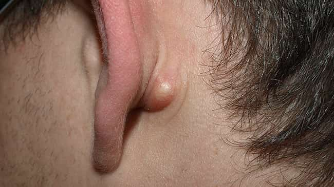
The Tumor Under the Ear
In this case, the child was suffering from a large tumor under her right ear. The cancer was about the size of a lemon at birth, and as she grew, it gradually increased in size and weight! In fact, according to her parents, the mass got so big that their daughter’s face changed shape, and she looked like a hamster!
The cause of such a large tumor is not known. Still, some potential causes may be an overactive pituitary gland leading to excessive production of Human Growth Hormones or an Overactive Thyroid Gland.
Another possible cause could be a combination of tumors affecting various glands such as the parathyroid, adrenal gland, and thymus. These tumors are called “Neoplasm,” which means new growth.
Neoplasms can occur anywhere in the body and are categorized by their tissue of origin, i.e., where they originate from, for example, bone, muscle, etc. This kind of tumor is called a “Hemangioma.” Since these tumors contain lots of blood vessels, they have lots of high-oxygenated blood, appearing bright red or purple.
Some hemangiomas such as this one may bleed with minor injuries such as rubbing against bedclothes or clothing. Generally, most do not produce any symptoms unless they grow huge and compress other tissues around them.
When tumors reach a size greater than 5 centimeters (2 inches) diameter, they can cause functional disturbances such as breathing difficulties, poor feeding, recurrent ear infections, etc. Therefore, doctors try to prevent further growth or reduce the size of hemangiomas with medication if possible to help avoid the need for surgery.
Surgery is usually undertaken when medication fails. The aim of surgery is not only to remove the tumor but also to correct any associated abnormalities such as facial distortion, which may have occurred due to the presence of a large mass under the skin. Other risks include bleeding and damage to surrounding structures such as nerve endings, resulting in numbness over part of the face following surgery.
The surgery for this child involved removing the tumor under general anesthesia. The surgery was performed by a senior maxillofacial surgeon in an operating theater in the hospital.
First, an incision was made behind her ear to expose the tumor, which had become very large and complex due to its size. The facial nerve, which runs just below where cancer lies, was observed during this procedure to avoid any injury if it got trapped between the tumor and surrounding structures.
The mass was dissected from other tissues to remove it without damaging structures, such as facial nerves or vessels running behind the ear. Some bleeding occurred, but it was easily controlled by applying a pressure bandage until hemostasis occurred (blood goes back to normal). The wound was left open to heal by secondary intention (without sutures) to avoid the risk of infection.
The child came back for review one week after surgery, and at that time, the wound had healed well, she was feeling refined, and her facial features were restored. She also had no signs of any bleeding or problems with facial nerve function.
Surgery is often necessary when tumors grow to a specific size, interfering with normal functioning. These tumors are usually benign (non-cancerous) but cause functional disturbances due to their bulk or location near essential nerves or vessels.
Surgery can be used not only as treatment but also as a preventive measure if it is anticipated to cause significant symptoms in the future, e.g., difficulties with breathing or swallowing.
Despite high-quality treatment and good medical care, some children may not respond to treatment, and they may develop complications such as Facial nerve injury, Bleeding following surgery, Infection of the wound, Trouble eating and taking enough nourishment in the long term due to problems chewing or swallowing Scarred skin resulting in cosmetic deformity
All these factors need to be considered before undertaking any open surgery. Still, if necessary, all possible efforts will be made at Children’s Hospital Los Angeles (CHLA) to provide the highest quality care so that our patients can enjoy a bright future.
lump under ear lobe behind the jaw bone:
if there is a lump under your ear lobe (behind the jaw bone), it means that you have thyroid gland swelling. This will be seen primarily in women undergoing heavy menstrual periods.
This could also be due to cysts, fluid-filled sacs containing watery fluid. These resting sacs are called hydatids and must be treated with surgery (unnecessary to remove all of them). But if they grow bigger, they should be removed by an operation.
However, this condition can also occur in men, primarily due to infection caused by staphylococcus aureus bacteria.
Treatment for men:
antibiotics or antibacterial drugs; however, treatment does not involve removal of the cyst
If you have a small lump that is not painful, you don’t need to worry about it. But if you have an infected cyst, it will be painful and filled with pus and may even burst open. The fluid from this abscess can spread throughout your face or neck, making it red, swollen, and sore.
A lump under the ear lobe could also be due to cancerous tumors called lymphoma, which form in the lymphatic system – part of the body’s immune defense network. Medical advice should always be sought as soon as possible because most cancers are treatable during their early stages.
Hard painless lump behind ear:
if you have a hard painless lump behind your ear, it may be due to an injury. The injury can result in a hematoma (a blood clot that forms under the skin). This is more common, especially if you play sports like wrestling, boxing, etc., it is unlikely that this condition would occur without any contact of some sort.
But suppose there’s no sign of trauma or injury. In that case, this could also be due to degenerative changes – meaning the normal process of aging where healthy tissues are replaced with fibrous tissue and calcium deposits (over time, these calcium deposits become more significant and harder). If you suspect this condition, it is best to consult your doctor.
If the lump behind your ear doesn’t get better over 4-6 weeks; if the swelling gets bigger or moves around from one place in your body to another; if there’s a redness in the area of the lump with pus coming out of it – then see a doctor for further treatment and tests.
Home remedies for a lump behind the ear:
1) hot fomentation:
you can do this by heating a towel in the microwave for 30 seconds and applying it to the lump behind your ear. Or, boil some water and hold that cloth (wet with warm water) over the affected area. This will help reduce the swelling and pain due to fluid buildup under your skin.
2) aloe vera gel:
aloe vera gel helps heal tissues faster; reduces pain, swelling, and redness after a wound. All you have to do is extract fresh aloe vera from a leaf of the aloe vera plant and rub it on the ear lobe. You can also use store-bought pure aloe vera gel – make sure that there are no preservatives in it.
3) turmeric paste:
turmeric has anti-inflammatory, antibacterial, and antioxidant properties, which help to reduce pain and swelling. All you have to do is mix some turmeric powder with warm water or castor oil (if your skin can tolerate this – castor oil helps to penetrate through the skin, so it reaches the hematoma more effectively). After mixing, apply to the affected area for 30 minutes, then wash off with plain water. Repeat this process three times a day for the best results.
4) onion juice:
onion juice contains anti-inflammatory substances which help to reduce swelling around the ear lobe area. All you have to do is take an onion and cut it in half. Now, take a fork and prick the onion – you can use as many pricks as you like (make sure to avoid the center of the onion where there’s a high concentration of sulfur).
After that, put this piece of onion on a plate and microwave for 30 seconds. Now apply the juice from the soft onion pieces on your swollen ear lobe area using a cotton ball or swab….and leave it on overnight. Chop up another half of an onion and repeat the process.
