What are The Causes of Uterine Cancer?
The following are the leading causes of uterine cancer:
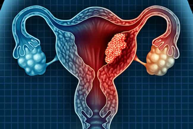
When cells begin to reproduce rapidly, this is called cancer. The development of tumors in the uterus may lead to uterine cancer. Cancerous growths start when healthy cells multiply quickly and crowd out other normal tissues. This is known as carcinogenesis or tumor formation.
While there are several reasons for carcinogenesis, two common causes are hereditary factors and environmental hazards. Both can be prevented by early detection with regular examinations by a physician specializing in detecting cancers like the gynecologist, family doctor, or general practitioner (GP).
Uterine cancer mainly results due to risk factors like:
Age – Risk increases after menopause; most cases occur in women over 50
Obesity – Overweight or obese people may be at greater risk of uterine cancer.
Prolonged use of oral contraceptive pills (OCPs) – Reports suggest that risks increase with extended periods of use, and the lowest stakes are seen after stopping pill use for ten years.
Smoking – One study found that smoking is a significant risk factor for endometrial cancer. Several studies have shown an association between heavy smokers (>20 cigarettes per day) and endometrial cancer, while some studies have associated light smoking (<20 cigarettes per day).
Drinking alcohol regularly – Drinking more than two drinks a day also increases the risk of uterine cancer.
Risk factors for uterine cancer include:
Family history of uterine or ovarian cancer
Undiagnosed vaginal bleeding
Age – Risk increases after menopause; most cases occur in women over 50
Obesity – Overweight or obese people may be at greater risk of uterine cancer.
advanced uterine cancer symptoms
how quickly do womb cancer spread
types of uterine cancer:
1)Endometrial cancer, also known as uterine cancer or cancer of the womb, is one of the main types of gynecologic cancers. Endometrial cancer forms in the tissues of the uterus (womb).
2)Uterine sarcoma:
Uterine sarcomas are rare and aggressive tumors that can develop in any muscle or connective tissue found within the walls of your uterus. This includes smooth muscle, which lines the uterine blood vessels and other structures, including fibroids.
It usually affects women ages 40 to 60 years old, although it’s possible for people younger than 40 or more aged than 60 to get uterine sarcoma. The symptoms vary depending on where you find the tumor.
3)Cervical cancer:
Cervical cancer forms in the tissues of your cervix (the lower part of the uterus that extends into the vagina). Cancerous cells sometimes develop from normal cells on the surface of the cervix. These changes take place slowly over the years, usually without any symptoms. However, these changes may eventually cause cervical cancer to develop.
4)Endometrial hyperplasia:
Endometrial hyperplasia is a precancerous condition in which the endometrium becomes enlarged and has more cells than usual. It’s not cancer or endometrial tissue growths (fibroids). But it does increase your risk of getting endometrial, so treatment should be considered.
5)Endometrial stromal sarcoma:
Endometrial stromal sarcoma is uncommon. It’s also difficult to detect because it usually doesn’t cause symptoms until the cancer is advanced. Women who develop endometrial stromal sarcoma are typically older than 45, and most cases occur in women age 60 or older.
6)Low malignant potential tumors (LMP tumors):
Some uterine growths that aren’t cancer but contain abnormal cells like endometrial polyps can develop into low malignant potential (LMP). These LMP tumors often produce estrogen, which helps feed the growth of other types of uterine cancer. This means that LMP tumors can lead to other uterine cancers.
uterine cancer stages:
The TNM staging system is used to describe the spread of cancer from where it started to other parts of the body. The earliest stage of uterine cancer is a stage I or IA when cells are found only in the inner lining of the uterus (endometrium) and have not grown into deeper layers.
Stage II uterine cancers have developed more profound into the tissue beneath the endometrium but haven’t spread outside the uterus. Stage III tumors have broken through these deeper layers and maybe grow into nearby tissues or organs, including structures inside your pelvis such as your colon, bladder, or rectum.
T-category defines primary tumor(tumor size):
T1 means that a tumor can’t be seen with a microscope but is big enough to be seen during surgery. T2 tumors are more prominent than T1 and can’t be removed by surgery without taking extra tissue from other parts of your body or removing the organ.
T-category defines primary tumor spread(tumor spread):
The letters T3, T4, and TNM describe tumor spread from where it first developed to nearby tissues.
T3 means that cancer has broken through the uterus wall into your inner pelvis or has grown into your cervix or vagina. This includes endometrial stromal sarcoma. Or it may have spread outside of your pelvis and lower abdomen to an area around those organs as on one side of your pelvis.
T4a indicates that cancer has invaded a nearby organ or tissue. T4b means distant metastasis (cancer that’s spread to a distant organ, such as the lungs).
uterine cancer bleeding pattern:
menstrual bleeding (during periods)
bleeding after menopause
uterine cancer frequency in males:
Male uterine cancer is rare. It’s called a testicular tumor in males after puberty and is almost always benign (not cancer).
uterine prolapse:
uterine prolapse happens when the uterus drops into or protrudes out of the vagina. The risk of developing uterine prolapse increases if you’ve had several pregnancies, significantly more than one vaginal delivery. However, uterine prolapse can occur without childbirth.
Uterus removal surgery:
Surgery to remove your uterus is called hysterectomy. Depending on your condition, you may have an abdominal hysterectomy or a vaginal hysterectomy. If only the cervix is affected by cancer, your doctor may do a radical hysterectomy and take out the upper part of your vagina, called a radical trachelectomy.
This surgery is only done if you are in good health; otherwise, with no complications to other organs.
Uterine cancer diagnosis:
Several tests can be used to diagnose uterine cancer. If you have symptoms or your doctor thinks you might have this condition, they may ask for one or more of these tests:
A pelvic exam helps find any abnormal tissue inside your uterus during a physical exam. Your doctor will insert one gloved finger into your vagina, which allows them to feel the shape and size of the uterus. A Pap smear may be taken simultaneously to examine cells from the cervix and vagina.
Transvaginal ultrasound :
An ultrasound uses sound waves and a computer to create images of structures inside your body. It can help determine whether there’s abnormal tissue growth in your uterus and surrounding areas. Ultrasound is used more often than any other imaging test because it doesn’t use radiation and is painless.
Hysteroscopy :
A hysteroscopy is an instrument that’s inserted through your vagina into your uterus for diagnostic purposes or surgery. An endometrial biopsy may be done during this procedure, depending on why you have it. Your doctor can also take small tissue samples (biopsies) from inside the uterus during a hysteroscopy.
Endometrial biopsy :
An endometrial biopsy is a procedure to remove samples of the uterine lining for testing. This test is usually done if you have abnormal bleeding from your vagina between periods or after menopause, especially if these conditions begin suddenly.
It may also diagnose uterine cancer before surgery or as a follow-up after treatment. The procedure involves inserting a thin needle through the wall of the vagina and into the uterus. Samples are taken from inside the uterus and checked for signs of cancer or other diseases under a microscope.
Preoperative tests :
In addition to those listed above, you’ll likely need several standard diagnostic tests before surgery to determine how advanced your disease is, how well your organs are functioning, and whether you’re healthy enough to have surgery. These tests may include blood counts, blood chemistries, kidney function tests, liver function tests, thyroid studies, cardiac stress testing (cardiac imaging), and an EKG.
Chest X-ray :
A chest x-ray is often taken before having surgery to remove the uterus. This can help determine whether there are signs of cancer in your lungs or other parts of your body that may not be near the pelvis.
Uterine cancer stages:
The most critical factor in treating uterine sarcoma is the diagnosis stage the disease has reached. Steps 0 through 4 are based on how far the tumor has spread beyond its original site within the uterus.
Stages 0 and 1 are often treated with surgery, while more advanced settings usually require additional treatments along with surgery.