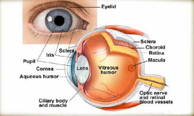
Eye Components
(1) cornea
(2) pupil
(3) iris (color of eyes)
(4) sclera (white part of the eye, surrounds the whole eye.)
The function of the human eye is to allow us to see. The main parts are:
(1) cornea
The cornea functions as a lens and protects the front of the eye. It is transparent and light passes through it (due to its transparency). Through the cornea, we can see clearly. Some people say that if we look at our corneas in a mirror they seem blue, but this is because the blood vessels show through more on them than they do on other parts of our bodies.
(2) pupil
The pupil (the black/grey bit) allows us to change how much light gets into our eyes. The pupil lets us control our vision by allowing only the right amount of light to focus on things around us. Light travels from within the eye towards the back part of it.
(3) iris (Colour of the eye)
The iris contains pigment that gives our eyes their color. It is sometimes called the pupil, although it isn’t a part of it. The colored part of the eye is called the iris. It looks like a small opening or hole in your eyeball. The size of this hole can be changed to control how much light comes into your eyes.
Myopia is usually caused by having too strong eyeballs, which makes focusing on near objects difficult due to its difficulty bending incoming light properly. Asians are more likely to be myopic than Caucasians.
Myopia can also lead to retinal detachment or glaucoma if untreated, leading to permanent blindness. People with myopia can wear glasses, contact lenses, or have surgery to correct their vision.
(4) sclera (The white part of the eye, surrounds the whole eye.)
The pigment in the eyes is brown-ish yellow and this is why our eyes are not blue or completely white. The color of your iris leans more towards different colors depending on what race you belong to.
It has been suggested from some research that Asians tend to have more brown pigment which makes their eyes appear browner than whites who have a higher concentration of yellow pigment in them. Brown also looks darker than yellow does, so appears black. So it’s a myth that people with dark eyes color look harder at things whereas light-eyed people look gentle.
A person’s eye color is determined by the amount of melanin in their iris. The more melanin you have, the darker your eyes will be.
People with lighter eyes tend to be short-sighted than people with dark eyes which is what causes them to wear glasses or contact lenses. Asians typically have a brownish-yellow color in their eyes and hence they are more likely to become short-sighted than whites who usually have blueish grey irises which makes them less likely to be myopic than Asians.
Also, darker pigmentation around light-colored eyes helps keep out stray lights that cause “halos” when driving at night compared to fair or pink-colored iris which does not help much in keeping out such extra lighting sources. This is the reason why my mother has her eyes checked for new lenses on an everyday basis. She is short-sighted and near-sighted than me even though I am an Asian too.
This also explains why many Asians will look at you with an evil eye if you stare or laugh at them because they are not used to light-colored eyes that often belong to Whites, Eurasians, or Mixed Races. Most of them are very protective of their kids have gray/blue/green/light brown eyes color because it means that they carry mostly Caucasian genes.
(5) optic nerve
The optic nerve carries signals from the retina to our brains so we can process what we see. It is sometimes called the second retina due to how it senses light differently.
The optic nerve of our eyes is like that of a computer. It is the connection between the computer’s screen (your eye) and your brain (your mind). A video signal (image) on a computer screen can be described as an array of dots. The light image on the screen is made up of millions of tiny dots, which you cannot see without looking at them closely.
eye parts and functions:
The eye is the organ that detects light and sends information to the brain. It has three main parts:
There are also five eyelids. The upper eyelid closes when you blink, just like your mouth’s upper lip closes when you sip through a straw. Note that in most animals (including humans), the eyes are on either side of the head so they can see what is coming towards them, allowing for an easier escape from danger.
By comparison, birds have their eyes placed at the front-center of their heads so they can better judge distances while flying (try it yourself!).
The pupil dilates (becomes larger) or contracts (becomes smaller). Pupils respond to changes in light levels by contracting or dilating. The pupil’s size also affects the amount of light that enters the eye. In a very bright room, your pupils become small to restrict the entry of light and to avoid damage.
In addition to these external parts, there are certain parts within the eyes to like:
retina (at the back), Iris (colored part), and cornea (clear front).
Retina: It is a membrane found in an inside layer of the eyeball; it contains cells that respond to light and transmit signals about what you’re looking at through the optic nerve to your brain.
The retina contains both rods and cones:
Rods help vision in dim lighting and provide peripheral vision because their receptive fields cover a wide area. Rods do not detect color. Rods, which are more light-sensitive than cones, contain a pigment that is sensitive to light called rhodopsin.
Rods work better in low light but do not give as sharp of an image as cones and provide less detail on images compared with cone vision. Rods also handle scotopic (night) vision, whereas cones deal with photopic (light) visual perception.
Cones tell your brain about the colors you see, as well as information on texture and shape. Cone cells respond only to fairly intense light sources. Cones make up the fovea of the retina – this area is responsible for central detailed high acuity vision – think about looking at a star or something close through a telescope.
Cones work better in bright light conditions, but they do not function as well at the edges of your vision. They provide more detailed information of objects you see compared with rods and are responsible for color vision. In cone cells, the pigment is called iodopsin (also found in rods).
The way cones sense color is by perceiving which wavelengths of light are reflected them by the object you’re looking at; this is known as trichromacy. The three types of cone cells respond differently to red, green, and blue light respectively giving you what we call full-color vision (or tetrachromacy).
functions of the eye:
Light enters the eye through the cornea.
The light then passes into the pupil (controlled by the iris), through the lens, and is focused onto the retina at the back of the eyeball.
Retina allows you to see images because it contains millions of nerve cells that react when hit with light, causing electrical impulses to be sent along the optic nerve to the brain where they are interpreted as sight. These impulses travel at about 186 miles per hour (300 kilometers per hour).
The eyelids protect the eyes by keeping out dust, dirt, and other foreign objects. The upper eyelid has an oily layer which is part of the tear film that lubricates the eye to prevent it from drying out.
The lachrymal gland also known as the tear gland or lacrimal gland is one of the cutaneous exocrine glands of the face. It produces tears, which keep the outer surface of your eyes moist. Tears also flow over your cornea each time you blink to keep it clean and moist.
Causes:
Lack or loss of vision can be caused by many reasons that include:
Diabetes mellitus Diabetic retinopathy Glaucoma CataractsKeratitisRetinal detachment Optic neuropathy Anterior ischemic optic neuropathy Posterior ischemic optic neuropathy Accommodative insufficiency Pseudoexfoliation syndrome
Diabetes retinopathy:
1) It is one of the main causes of blindness in adults. Diabetes can damage blood vessels in the retina.
If not treated, it may cause your vision to become blurry or black spots to appear in your sight.
2) Glaucoma. It is a disease where increased pressure inside the eye leads to progressive loss of vision due to the death of nerve cells at the back of the eyes. Glaucoma results from increased intraocular pressure (IOP). High IOP put direct stress on the optic nerve.
Normal IOP is 10-21mmHg, while glaucoma can be diagnosed when it is 22mmHg or more. In addition to high pressure, the death of nerve cells may also result from bleeding in the eye or optic nerve damage. If taken care of on time glaucoma can be treated very well by Laser surgery and medications.
3) Cataracts. It occurs due to the aging process where clarity of lens decreases gradually resulting in vision changes. It affects both eyes equally.
4) Keratitis. It is an inflammation or infection that damages your cornea which can lead to scarring, pain, light sensitivity, and blurred vision.
5) Retinal detachment. When the retina separates from the choroid, then it is called retinal detachment.