Physiology of Anaphylaxis
anaphylaxis is an immediate hypersensitivity reaction that occurs when exposure to an allergen triggers the release of mast cells and histamines, thereby leading to systemic symptoms.
Anaphylactic reactions are often caused by food allergies but can be triggered by insect stings, exposure to medications, or latex proteins. Anaphylactoid reactions are similar in symptomology but do not involve true antigen-IgE mediated sensitization.
Diagnosis & management:
patients with suspected anaphylaxis must present immediately for evaluation after exposure, irrespective of the severity of the reaction, especially if one of the six “red flag” signs (throat tightness; swelling; shortness of breath; sizing; unconsciousness; not breathing) is present or if there are cardiovascular changes (hypotension, tachycardia). Treatment includes epinephrine, antihistamines, and steroids. Patients should be observed for at least four hours after the first dose of epinephrine to ensure the resolution of symptoms.
Risk factors:
the risk factors for anaphylaxis include allergy to peanuts, tree nuts, milk, egg white, fish; asthma; aspirin intolerance; bee sting allergies; some autoimmune conditions like lupus erythematosus (SLE), rheumatoid arthritis (RA), autoimmune thyroid disease.
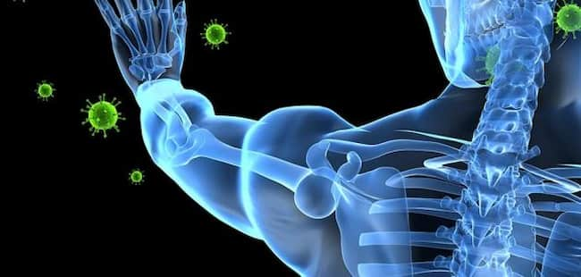
Anatomy & pathophysiology:
mast cells are part of the body’s standard defense system against foreign substances called antigens. Mast cells reside in most tissues but are found mainly in the skin, lungs, and intestines.
When an antigen enters the body that the immune system recognizes as potentially harmful, it combines with immunoglobulin E (IgE), a portion of which is attached to the surface of mast cells at sites called receptors.
The IgE molecules guide these receptors like radar:
they help activate the mast cell when an allergen or toxin binds to one of its antennae-like ends. This combination triggers a series of responses inside the cell; if enough IgE attaches to multiple mast cells and their antennas point toward each other, a domino effect can occur. The activated mast cells secrete chemicals such as histamine that promote inflammation and lead to allergic reactions.
Anaphylaxis:
Anaphylaxis occurs when mast cells release massive amounts of histamine and other chemicals into the bloodstream all at once after an allergen triggers their activation.
This response is often rapid in onset and potentially life-threatening; it stimulates smooth muscles throughout the body, including those in vessels (causing them to constrict) and airways (causing them to spasm).
The degree of constriction or spasm relates directly to the severity of anaphylaxis: if these muscles remain relaxed, blood vessels dilate appropriately, and breathing continues without difficulty; but if they contract too much, airflow through the lungs can be compromised, resulting in stuff. Other effects include low blood pressure, nausea, vomiting, diarrhea, and abdominal pain.
Anaphylactic shock:
It is the most severe manifestation of anaphylaxis, resulting when blood pressure decreases and blood supply to vital organs is substantially reduced.
Another cause:
several other conditions mimic an allergic reaction (and vice versa) or produce symptoms that overlap with those of anaphylaxis; these include basophilic leukemia; eosinophilic esophagitis; mastocytosis; angioedema; food poisoning (e.g., Staphylococcus aureus); scombroid (histamine poisoning from fish); hereditary angioneurotic edema; Herpes simplex infection; Kawasaki disease; and hyperventilation.
Diagnostic criteria:
1. Acute onset of an illness (minutes to several hours) with skin, mucosal tissue involvement, or both (e.g., generalized hives, pruritus or flushing, swollen lips-tongue-uvula).
2. Respiratory compromise (e.g., dyspnea, bronchospasm, stridor, laryngeal edema) and reduced blood pressure or associated symptoms of end-organ dysfunction (e.g., angioedema; hypotonia; nausea; vomiting; diarrhea).
3. Exposure to a likely allergen that can be confirmed by clinical history.
4. Symptoms resolve after the onset of anaphylaxis.
5. Onset of symptoms is rapid after exposure to a likely allergen (minutes to several hours).
6. Exposure to an allergen capable of producing systemic allergic reactions, including anaphylaxis, in patients with atopy and prior generation of antigen-specific IgE antibodies.
Management & treatment:
treatment may include supportive care, such as securing the airway and monitoring for hypotension; treating the allergic reaction with an epinephrine injection if indicated; and administering intravenous fluids. Additional drugs, such as steroids and H1 blockers (e.g., diphenhydramine), can treat more severe anaphylaxis manifestations.
Treatment:
Epinephrine is the most critical medication in treating anaphylaxis with a single injection providing rapid response with a relatively quick return of blood pressure, improvement in respiratory symptoms, and rarely minor cardiovascular changes.
Those exposed to the allergen again should have injectable epinephrine available for self-administration at home or school/work upon recognizing early signs and symptoms that may progress rapidly to anaphylaxis. Injectable epinephrine can also be given intravenously by professional teams, but this must be careful while monitoring vital signs and providing other life-supporting care.
The patient with mild anaphylaxis symptoms that have prompt resolution may be treated with self-injectable epinephrine if these are more severe or do not resolve promptly.
Patients with recurrent or persistent anaphylaxis should have injectable epinephrine immediately available for self-administration by their caregivers at home, school and work upon recognizing early signs and symptoms that may progress rapidly to anaphylaxis.
Pathophysiology of an allergic reaction:
An allergy is a hypersensitivity disorder of the immune system. This means that the body’s immune system, for unknown reasons, overreacts to substances in the environment that are generally not harmful (allergens).
Most people know exactly what they are allergic to; however, some suffer from allergies without knowing it. For example, hay fever (allergic rhinitis) and asthma can develop gradually with repeated exposure to allergens which provoke symptoms. Allergic reactions may be caused by food products, insect stings, medications, and latex.
An allergen stimulates an antibody called Immunoglobulin E (IgE), which binds to mast cells lining the respiratory passages or surface. The binding causes the release of histamine and other chemicals (mediators), which produce allergy symptoms.
Histamine causes blood vessels to become “leaky,” allowing fluid to escape into tissues, causing the nasal mucous membranes to become swollen and inflamed. Other mediators increase mucus production and cause smooth muscle contractions, resulting in a runny nose and other symptoms.
The immune system:
The immune system is composed of several distinct cellular and soluble components, all aimed at making our bodies inhospitable environments for pathogenic microorganisms such as parasites, fungi, viruses, or bacteria by using specialized cells to identify them as foreign substances (“non-self”) and neutralize them as efficiently as possible.
However, this same system sometimes fails to discern among harmless substances, standard components of the body itself, non-threatening parasites, or fungi and targets them as foreign or harmful. This results in allergies.
Four stages of anaphylaxis:
The process of anaphylaxis has been divided into four stages, each with distinct pathophysiology.
Anaphylactoid reactions lack the IgE involvement that is characteristic of true anaphylaxis. In this form, what begins as normal immune system responses to a stimulus results in a massive release of histamine and other pro-inflammatory mediators from mast cells and basophils.
Symptoms usually begin within minutes after exposure due to a direct effect on mast cells/basophils or a secondary release from damaged tissue by cytokines or toxic mediators.
Systemic symptoms characterize the second stage because of the access of substances via venous blood spreading throughout the body; it typically occurs 5-30 minutes after exposure to the allergen.
The third stage, occurring minutes to hours after the second phase, is characterized by angioedema and urticaria in addition to hypotension and vascular collapse.
The fourth stage results from mediator-induced tissue damage; it typically occurs 2-12 hours after exposure when cytokines induce inflammation and vasodilation, permanently altering tissues if untreated. This phase may also be called late anaphylaxis or biphasic reaction.
an acute allergic reaction:
An acute allergic reaction (also referred to as type 1 hypersensitivity) is mediated by mast cells and basophils upon reexposure to the same antigen that stimulated their original production. Thus, for example, An individual stung by a bee who has never before experienced the reaction might notice itching at the site of the sting and no other symptoms.
However, should this individual be stung by another bee in that area, they may experience redness, swelling, itching, and pain. This is because IgE antibodies have already bound to mast cells in the common areas (such as skin or eyes), which triggers them to release histamine into these tissues. In addition, histamine causes itchiness within seconds due to its vasodilation properties.
causes of anaphylaxis:
The causes of anaphylaxis are widespread:
Plants Food products Pets Medications Latex Physical stimuli (e.g., heat or cold) May occur without prior exposure to the allergen, in which case, it is called idiopathic (i.e., unknown cause).
Symptoms:
Symptoms usually begin within minutes after exposure due to a direct effect on mast cells/basophils or a secondary release from damaged tissue by cytokines or toxic mediators.
They can vary according to the type of reaction being experienced. Types of hypersensitivity reactions are:-
IgE-mediated reactions, cell-mediated allergic reaction, immediate contact reaction, phagocytic cell reaction, delayed-type hypersensitivity reaction, cytotoxic, and T lymphocyte reaction.
It generally takes 15 to 30 minutes for symptoms of acute anaphylaxis to appear after exposure to the provoking allergen.
Cutaneous features characterize the first stage (e.g., flushing, urticaria), respiratory features (shortness of breath, bronchospasm), gastrointestinal features (nausea, vomiting), and cardiovascular collapse (“shock”).
During this first phase, mast cells degranulate and release vasoactive amines such as histamine, which induce systemic symptoms. Histamine acts on smooth muscle, causing bronchoconstriction; blood vessels are more absorbent, which causes hypotension; intestinal peristalsis slows or stops.
The second stage is characterized by laryngeal edema (swelling of the larynx), generalized wheezing, and reduced blood pressure. If the epinephrine effect is insufficient to compensate for the vasodilatation caused by histamine, there may be a drop in blood pressure, leading to brain hypoperfusion.
The third stage, occurring minutes to hours after the second phase, is characterized by angioedema and urticaria in addition to hypotension and vascular collapse. Vasodilation contributes to hypotension; however, it also causes increased cardiac output, leading to heart attacks or strokes if not treated.